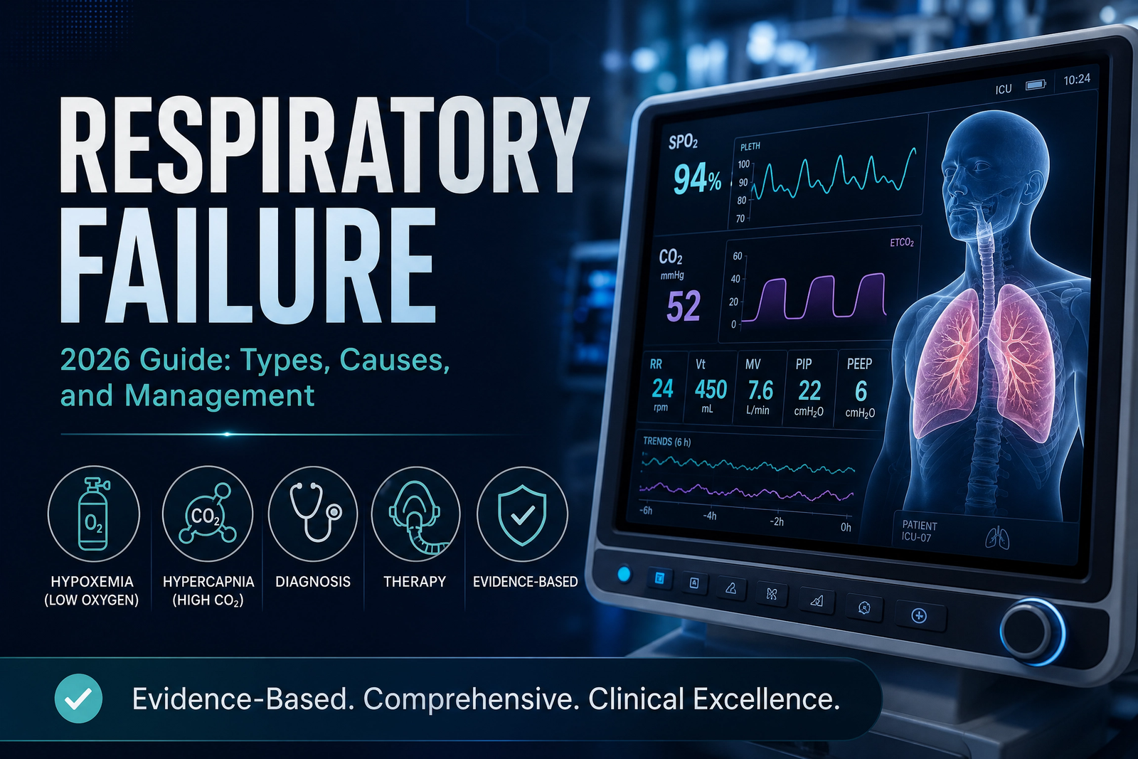
Respiratory Failure: Types, Causes, Diagnosis, and Evidence-Based Management
A comprehensive, peer-reviewed clinical resource on acute and chronic respiratory failure — from pathophysiology to advanced ventilatory support
I. Respiratory Failure: Definition and Clinical Significance
Respiratory failure is a life-threatening condition in which the respiratory system fails to maintain adequate gas exchange, resulting in either hypoxemia (PaO₂ <60 mmHg), hypercapnia (PaCO₂ >50 mmHg), or both. It is the final common pathway of numerous pulmonary, cardiac, neuromuscular, and central nervous system disorders, and is the leading cause of intensive care unit (ICU) admission worldwide.
Respiratory failure is classified by onset (acute vs. chronic) and by the primary gas exchange abnormality (Type I: hypoxemic vs. Type II: hypercapnic). This classification guides diagnostic testing, treatment selection, and prognosis. Acute respiratory failure is a medical emergency requiring immediate intervention, while chronic respiratory failure develops gradually over months to years and may be managed with outpatient strategies including non-invasive ventilation and pulmonary rehabilitation.
• 2-3 million ICU admissions annually in the US for respiratory failure
• Leading cause of ICU admission worldwide
• 30-50% hospital mortality for acute respiratory failure requiring mechanical ventilation
• 1-year mortality for chronic respiratory failure: 20-40%
• ARDS accounts for 10% of ICU admissions and 25% of mechanical ventilation days
II. Type I vs. Type II Respiratory Failure: The Fundamental Distinction
| Feature | Type I (Hypoxemic) | Type II (Hypercapnic) |
|---|---|---|
| Definition | PaO₂ <60 mmHg, normal or low PaCO₂ | PaCO₂ >50 mmHg, low or normal PaO₂ |
| Primary Defect | Impaired oxygenation | Impaired ventilation |
| Pathophysiology | V/Q mismatch, shunt, diffusion impairment, low FiO₂ | Hypoventilation, neuromuscular weakness, central depression, chest wall restriction |
| Typical Causes | ARDS, pneumonia, pulmonary edema, pulmonary embolism, interstitial lung disease | COPD, neuromuscular disease (ALS, Guillain-Barré), opioid overdose, obesity hypoventilation syndrome, kyphoscoliosis |
| Response to O₂ | Improves (except shunt) | Improves PaO₂ but may worsen CO₂ retention (in susceptible patients) |
| Treatment | Supplemental oxygen, treat underlying cause, manage V/Q mismatch | Improve ventilation (NIV/BiPAP, mechanical ventilation), treat underlying cause |
| Prognosis | Variable; depends on underlying cause | Variable; depends on reversibility of underlying cause |
The Alveolar-Arterial (A-a) Gradient in Type I Failure
In Type I respiratory failure, the A-a gradient (PAO₂ - PaO₂) is typically elevated (>20 mmHg on room air, increasing with age). This indicates intrinsic lung pathology (V/Q mismatch, shunt, or diffusion impairment). A normal A-a gradient with hypoxemia suggests hypoventilation (Type II) or low inspired oxygen (high altitude). Calculating the A-a gradient is essential for distinguishing pulmonary from extrapulmonary causes.
III. Causes and Risk Factors of Respiratory Failure
Pulmonary Causes
Chronic Obstructive Pulmonary Disease (COPD): The most common cause of chronic Type II respiratory failure. Progressive airflow obstruction, hyperinflation, and respiratory muscle dysfunction culminate in CO₂ retention. Acute exacerbations (infections, air pollution) can precipitate acute-on-chronic respiratory failure requiring NIV or mechanical ventilation.
Pneumonia (CAP, HAP, VAP): Infection-induced inflammation, alveolar filling, and V/Q mismatch cause Type I respiratory failure. Severe pneumonia may progress to ARDS. Mortality increases with severity and delayed antibiotic administration.
Acute Respiratory Distress Syndrome (ARDS): Diffuse alveolar damage from sepsis, pneumonia, trauma, or pancreatitis. Characterized by bilateral opacities on CXR, non-cardiogenic pulmonary edema, and PaO₂/FiO₂ ratio ≤300. Lung-protective ventilation reduces mortality.
Pulmonary Edema (Cardiogenic): Elevated pulmonary capillary wedge pressure (PCWP) >18 mmHg from left ventricular dysfunction. Interstitial and alveolar fluid impairs gas exchange, causing Type I respiratory failure. Treatment includes diuretics, vasodilators, and inotropic support.
Pulmonary Embolism (PE): Acute obstruction of pulmonary arterial circulation creates dead space ventilation, V/Q mismatch, and right heart strain. Massive PE causes hemodynamic instability and Type I respiratory failure. Treatment includes anticoagulation and, in massive cases, thrombolysis or embolectomy.
Neuromuscular Causes
Amyotrophic Lateral Sclerosis (ALS): Progressive motor neuron degeneration leads to respiratory muscle weakness, impaired cough, and Type II respiratory failure. Nocturnal NIV improves survival and quality of life.
Guillain-Barré Syndrome (GBS): Acute demyelinating polyneuropathy causes ascending paralysis affecting respiratory muscles. Up to 20-30% of GBS patients require mechanical ventilation during the acute phase.
Myasthenia Gravis: Autoimmune neuromuscular junction blockade causes fluctuating weakness, affecting extraocular, bulbar, and respiratory muscles. Crisis management includes IVIG/plasmapheresis and ventilatory support.
Central Nervous System Causes
Opioid Overdose: Central respiratory depression through mu-opioid receptor stimulation in the brainstem. Naloxone reversal, bag-mask ventilation, and support until drug metabolism are critical.
Brainstem Stroke or Hemorrhage: Disruption of respiratory centers in the medulla (pre-Bötzinger complex) causes failure of central respiratory drive. Mechanical ventilation is required until neural recovery or determination.
Chest Wall and Extrapulmonary Causes
Obesity Hypoventilation Syndrome (OHS): Excess chest wall mass increases work of breathing, causing chronic hypoventilation and Type II respiratory failure. Weight loss and NIV are first-line treatments.
Kyphoscoliosis: Severe thoracic deformity restricts lung expansion, increasing work of breathing and predisposing to Type II respiratory failure.
COPD patients with chronic hypercapnia are at risk of oxygen-induced hypercapnia when high-concentration oxygen is administered. This occurs through three mechanisms: reduced hypoxic ventilatory drive, the Haldane effect (CO₂ displacement from hemoglobin), and reduced hypoxic pulmonary vasoconstriction (worsening V/Q mismatch). Target SpO₂ 88-92% in these patients.
IV. Clinical Presentation of Respiratory Failure
The clinical presentation of respiratory failure depends on severity, acuity, and underlying cause.
Early Signs (Compensatory Phase)
- Tachypnea: Respiratory rate >20 breaths/min as the body attempts to increase minute ventilation
- Tachycardia: Heart rate >100 bpm to augment oxygen delivery
- Diaphoresis: Sympathetic activation from hypoxemia/hypercapnia
- Restlessness and anxiety: Cerebral hypoxia and CO₂-mediated stimulation
- Headache: CO₂-mediated cerebral vasodilation
Late Signs (Decompensation)
- Cyanosis: Bluish discoloration of lips, tongue, and fingertips indicates deoxygenated hemoglobin >5 g/dL (late sign)
- Altered mental status: Confusion, agitation, somnolence, obtundation, or coma
- Asterixis: Flapping tremor of outstretched hands (CO₂ encephalopathy)
- Papilledema: Optic disc swelling from elevated intracranial pressure (chronic hypercapnia)
- Accessory muscle use: Sternocleidomastoid, scalene, and intercostal muscle recruitment indicates increased work of breathing
- Paradoxical breathing: Abdominal wall moves inward during inspiration (diaphragm fatigue)
• Severe shortness of breath at rest
• Inability to speak full sentences
• Blue lips or fingernails (cyanosis)
• Confusion, extreme drowsiness, or inability to stay awake
• Chest pain with breathing difficulty
• Rapid breathing (>30 breaths/min) with accessory muscle use
V. Diagnostic Approach to Respiratory Failure
Arterial Blood Gas (ABG) — The Gold Standard
ABG analysis is the diagnostic cornerstone for respiratory failure.
Normal values: PaO₂ 80-100 mmHg (95-100% SpO₂), PaCO₂ 35-45 mmHg, pH 7.35-7.45, HCO₃⁻ 22-26 mEq/L.
Type I (Hypoxemic) Failure: PaO₂ <60 mmHg, PaCO₂ normal or low, pH normal or alkalotic (hyperventilation).
Type II (Hypercapnic) Failure: PaCO₂ >50 mmHg, pH low (acute) or normal (chronic compensation), HCO₃⁻ normal (acute) or elevated (chronic renal compensation).
Interpretation of ABG in Acute vs. Chronic Respiratory Failure
Acute hypercapnia: PaCO₂ >50 mmHg, pH <7.35, HCO₃⁻ normal (22-26). No time for renal compensation.
Chronic hypercapnia: PaCO₂ >50 mmHg, pH 7.35-7.40, HCO₃⁻ elevated (>26). Renal compensation has occurred over 48-72 hours.
Acute-on-chronic: PaCO₂ elevated above patient's baseline, pH decline >0.05 from compensated level. This is a medical emergency requiring urgent intervention.
Additional Diagnostic Testing
- Chest radiography: Detects pneumonia, atelectasis, pneumothorax, pulmonary edema, or infiltrates
- Computed tomography (CT): Superior detail for ILD, PE, or cavitary lesions
- Pulmonary function tests (PFTs): Spirometry, lung volumes, DLCO identify restrictive or obstructive patterns
- Echocardiography: Assesses cardiac function and excludes cardiogenic pulmonary edema
- Complete blood count (CBC): Identifies anemia (reduced oxygen-carrying capacity), infection (leukocytosis), polycythemia (chronic hypoxemia)
- Electrocardiogram (ECG): Arrhythmias, right ventricular hypertrophy (cor pulmonale)
- Serum biomarkers: Procalcitonin (PCT) for bacterial infection; BNP/NT-proBNP for cardiac failure
VI. Evidence-Based Treatment of Respiratory Failure
Controlled Oxygen Therapy
For Type I (hypoxemic) respiratory failure, oxygen is the first-line therapy. Target SpO₂ 94-98% for most patients. For Type II (hypercapnic) respiratory failure, target SpO₂ 88-92% to avoid oxygen-induced hypercapnia. Devices include nasal cannula (1-6 L/min), face mask (5-10 L/min), Venturi mask (precise FiO₂), and high-flow nasal cannula (HFNC) up to 60 L/min.
Non-Invasive Ventilation (NIV/BiPAP)
NIV via bilevel positive airway pressure (BiPAP) is first-line for acute Type II respiratory failure with pH 7.25-7.35, respiratory rate >24, and preserved consciousness. It reduces intubation rates by 35-50% and mortality in COPD exacerbations. It is also indicated for acute cardiogenic pulmonary edema and as a bridge to invasive ventilation.
Typical BiPAP settings: IPAP 10-14 cm H₂O, EPAP 4-6 cm H₂O, backup rate 12-14 breaths/min. Titrate to clinical response (improved pH, reduced respiratory rate, decreased PaCO₂).
Invasive Mechanical Ventilation
Indications for endotracheal intubation and mechanical ventilation include:
- Failed NIV (pH <7.25 despite BiPAP, worsening mental status)
- Severe hypoxemia (PaO₂ <60 mmHg on high-flow O₂ or FiO₂ >0.6)
- Respiratory arrest or apnea
- Inability to protect airway (aspiration risk, decreased GCS)
- Severe encephalopathy or agitation
- Hemodynamic instability
- Excessive work of breathing with impending respiratory exhaustion
Lung-protective ventilation principles:
- Tidal volume: 6-8 mL/kg predicted body weight (PBW)
- Plateau pressure ≤30 cm H₂O
- PEEP 5-15 cm H₂O (higher in ARDS)
- Respiratory rate 20-30 breaths/min (permissive hypercapnia acceptable)
- FiO₂ titrated to SpO₂ 88-95% (or 94-98% for non-COPD)
Landmark clinical trials (ARDSNet 2000) established that lung-protective ventilation with 6 mL/kg PBW tidal volume and plateau pressure ≤30 cm H₂O reduces mortality in ARDS from 40% to 31% (absolute risk reduction 9%). These principles now apply broadly to all patients with acute respiratory failure, regardless of ARDS diagnosis.
Treatment of Underlying Causes
Specific treatments targeting the underlying condition are essential:
- Antibiotics for pneumonia or sepsis
- Bronchodilators and corticosteroids for COPD/asthma exacerbations
- Diuretics for cardiogenic pulmonary edema
- Anticoagulation for pulmonary embolism
- Naloxone for opioid overdose
- IVIG/plasmapheresis for Guillain-Barré or myasthenia gravis
- Antifibrotic therapy (pirfenidone, nintedanib) for progressive fibrosing ILD
VII. Prognosis and Complications of Respiratory Failure
Mortality and Survival
Hospital mortality for acute respiratory failure requiring mechanical ventilation ranges from 30-50% depending on underlying cause, severity, and patient comorbidities. ARDS carries 30-45% mortality. COPD exacerbations with hypercapnic respiratory failure have 10-20% in-hospital mortality. Long-term survival after hospital discharge for ARDS is approximately 80% at 1 year, 70% at 3 years.
Complications of Respiratory Failure
- Ventilator-Associated Pneumonia (VAP): Occurs in 15-25% of mechanically ventilated patients. Prevention: head-of-bed elevation 30-45°, daily sedation interruption, oral chlorhexidine, subglottic suctioning.
- Barotrauma: Pneumothorax, pneumomediastinum, subcutaneous emphysema from high airway pressures.
- Ventilator-Induced Lung Injury (VILI): Volutrauma, atelectrauma, and biotrauma from excessive tidal volumes and pressures.
- ICU-Acquired Weakness: Myopathy and neuropathy from critical illness, inflammation, and immobility.
- Post-Intensive Care Syndrome (PICS): Cognitive, psychiatric, and physical impairments persisting after ICU discharge. Affects 30-50% of survivors.
VIII. Interactive Clinical Tools
🫁 Respiratory Failure Risk Assessment Calculator
This validated screening tool evaluates risk factors for respiratory failure based on clinical parameters and underlying conditions.
🩸 ABG Respiratory Failure Classifier
Enter arterial blood gas values to determine if you meet criteria for Type I or Type II respiratory failure.
Support Respiratory Health and Lung Function
Targeted nutritional support — including N-acetylcysteine (NAC), vitamin D, and antioxidant formulations — may help support respiratory health in individuals with chronic lung conditions. Consult your physician before starting any supplement regimen.
Explore Respiratory Support Formulas →These statements have not been evaluated by the FDA. This product is not intended to diagnose, treat, cure, or prevent any disease. Individual results vary.
IX. Frequently Asked Questions
Respiratory failure is a condition where the respiratory system fails to maintain adequate gas exchange, resulting in either low oxygen (PaO₂ <60 mmHg), high carbon dioxide (PaCO₂ >50 mmHg), or both. It is classified as acute or chronic based on onset and severity.
Type I (hypoxemic) respiratory failure is characterized by low PaO₂ (<60 mmHg) with normal or low PaCO₂. It results from V/Q mismatch, shunt, or diffusion impairment (ARDS, pneumonia, pulmonary edema). Type II (hypercapnic) respiratory failure is characterized by elevated PaCO₂ (>50 mmHg) with low or normal PaO₂. It results from hypoventilation (COPD, neuromuscular disease, central depression).
Causes include lung disease (COPD, asthma, pneumonia, ARDS, pulmonary fibrosis), neuromuscular disorders (ALS, Guillain-Barré, myasthenia gravis), central nervous system depression (opioids, brainstem stroke), chest wall restriction (kyphoscoliosis, obesity hypoventilation syndrome), and extrapulmonary causes (sepsis, heart failure, pulmonary embolism).
Diagnosis requires arterial blood gas (ABG) analysis showing PaO₂ <60 mmHg and/or PaCO₂ >50 mmHg. Additional tests include chest imaging (CXR/CT), pulmonary function tests (PFTs), echocardiography, and laboratory studies. Clinical signs include tachypnea, accessory muscle use, cyanosis, and altered mental status.
Treatment depends on severity and underlying cause. Options include: controlled oxygen therapy (Type I), non-invasive ventilation (BiPAP/NIV) for Type II, invasive mechanical ventilation (endotracheal intubation), treatment of underlying cause (antibiotics, bronchodilators, diuretics), and supportive care (pulmonary rehabilitation, nutrition).
Prognosis depends on severity, underlying cause, and comorbidities. Acute respiratory failure requiring mechanical ventilation has hospital mortality of 30-50%. Chronic respiratory failure (COPD, neuromuscular) has variable prognosis, with 1-year mortality of 20-40%. Early recognition and treatment improve outcomes.
Indications for mechanical ventilation include: failed NIV, severe hypoxemia (PaO₂ <60 mmHg on high-flow O₂), hypercapnia with pH <7.25, respiratory arrest, severe encephalopathy, inability to protect airway, hemodynamic instability, and excessive work of breathing. Lung-protective ventilation (tidal volume 6 mL/kg PBW, plateau pressure ≤30 cmH₂O) reduces mortality in ARDS.
Prevention strategies include: smoking cessation, vaccination (influenza, pneumococcal, COVID-19), optimal management of chronic respiratory diseases (COPD, asthma), early treatment of respiratory infections, avoiding respiratory depressant medications (opioids, benzodiazepines), and pulmonary rehabilitation for chronic lung disease.
Create an Optimal Recovery Environment
Proper sleep positioning and ergonomic sleep systems support respiratory mechanics and optimize breathing during rest. Maintain proper head and neck alignment to reduce respiratory effort and improve sleep quality.
Discover Ergonomic Sleep Solutions →These statements have not been evaluated by the FDA. Individual results vary. Always consult your physician.
Leave a Reply