
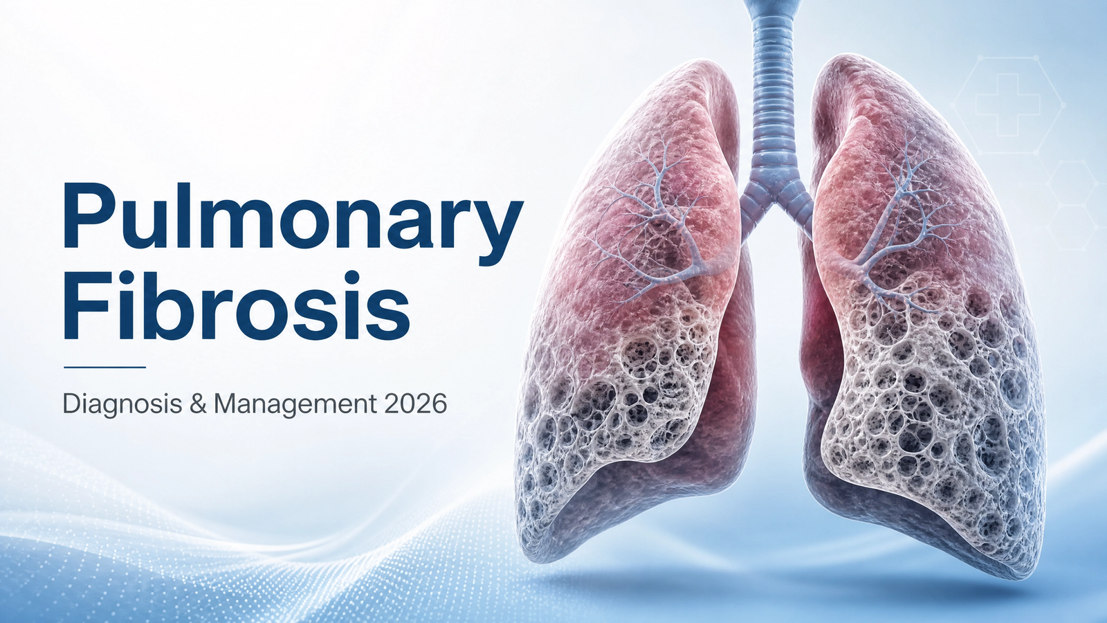
Pulmonary Fibrosis: Pathophysiology, Clinical Classification, Diagnosis, and Evidence-Based Management
A comprehensive, peer-reviewed clinical resource on interstitial lung disease — from molecular fibroblast activation to multidisciplinary diagnostic consensus and antifibrotic therapy
I. Introduction: The Global Burden of Pulmonary Fibrosis
Pulmonary fibrosis — progressive scarring of the lung parenchyma — represents one of the most challenging and fatal interstitial lung diseases (ILDs), with a median survival of 3-5 years from diagnosis in its most common form, idiopathic pulmonary fibrosis (IPF). The condition affects approximately 3 million people worldwide, with an estimated 100,000 Americans currently living with IPF. Incidence rises dramatically with age, peaking between 60-75 years, with a slight male predominance (1.5:1). Despite being classified as a rare disease, the global burden is substantial, and survival rates have only modestly improved with the introduction of antifibrotic therapy.
Unlike infectious or malignant lung diseases, pulmonary fibrosis is characterized by aberrant wound healing: the lung's normal repair mechanisms become dysregulated, leading to excessive deposition of extracellular matrix, progressive distortion of alveolar architecture, and ultimately, respiratory failure. The economic burden is immense — direct healthcare costs for IPF in the United States exceed $3 billion annually, and the indirect costs of disability and lost productivity are even higher.
• 3 million people affected globally with pulmonary fibrosis
• 100,000 Americans living with IPF
• 30,000-40,000 new IPF cases diagnosed annually in the US
• Median survival: 3-5 years from diagnosis (IPF)
• Peak incidence: 60-75 years (male predominance 1.5:1)
• 5-year mortality: 40-50% (higher than many cancers)
II. Molecular Pathophysiology: The Fibrotic Cascade
Pulmonary fibrosis results from a complex interplay of genetic susceptibility, environmental triggers, and aberrant cellular signaling. The final common pathway involves activation of fibroblasts into myofibroblasts, excessive deposition of extracellular matrix (collagen, fibronectin), and progressive destruction of alveolar architecture.
Epithelial injury and activation: The initiating event in pulmonary fibrosis is believed to be recurrent micro-injury to the alveolar epithelium. This injury triggers release of damage-associated molecular patterns (DAMPs) and alarmins (HMGB1, IL-33, IL-25), activating the innate immune system. Senescent alveolar epithelial cells (type II pneumocytes) accumulate and secrete pro-fibrotic mediators via the senescence-associated secretory phenotype (SASP).
TGF-β signaling: The master regulator: Transforming growth factor-beta (TGF-β) is the central mediator of fibrogenesis. Released from the extracellular matrix by proteolytic cleavage (mediated by integrins and matrix metalloproteinases), TGF-β binds to its receptor (TGF-βRII), phosphorylating SMAD2/3, which translocates to the nucleus and activates transcription of pro-fibrotic genes (collagen, fibronectin, α-SMA). The TGF-β pathway is activated in all forms of pulmonary fibrosis and represents a key therapeutic target.
Fibroblast-to-myofibroblast differentiation: Under TGF-β stimulation, resident fibroblasts and circulating fibrocytes differentiate into α-smooth muscle actin (α-SMA)-expressing myofibroblasts. These cells are the primary effector cells of fibrosis, depositing extracellular matrix at sites of injury. Myofibroblasts are resistant to apoptosis, enabling persistent fibrotic activity.
Extracellular matrix deposition and alveolar obliteration: Excessive collagen deposition (primarily types I and III) progressively replaces normal alveolar architecture. The characteristic "honeycombing" pattern on HRCT corresponds to dilated, fibrotic airspaces lined with bronchiolar epithelium. As fibrosis progresses, capillary networks are obliterated, creating ventilation-perfusion mismatch and progressive hypoxemia.
The MUC5B promoter polymorphism (rs35705950) is the most common genetic risk factor for sporadic IPF, present in 30-50% of patients. Mutations in telomere-related genes (TERT, TERC, PARN, RTEL1) account for 15-20% of familial PF. Surfactant protein gene mutations (SFTPC, SFTPA2) and rare variants in genes involved in host defense (TOLLIP, IL1RN) also increase susceptibility. Genetic testing is recommended for familial PF and early-onset disease.
III. Clinical Classification of Pulmonary Fibrosis
Pulmonary fibrosis encompasses a heterogeneous group of ILDs classified by etiology and histopathological pattern.
| Category | Specific Diagnosis | Key Features |
|---|---|---|
| Idiopathic Interstitial Pneumonias | Idiopathic Pulmonary Fibrosis (IPF) | UIP pattern on HRCT/histology; progressive fibrosis; no identifiable cause |
| Non-Specific Interstitial Pneumonia (NSIP) | Cellular or fibrotic NSIP; associated with CTD; better prognosis than IPF | |
| Cryptogenic Organizing Pneumonia (COP) | Bronchiolar plugs; steroid-responsive; migratory infiltrates | |
| Connective Tissue Disease-Associated ILD | Rheumatoid Arthritis (RA-ILD) | UIP or NSIP pattern; seropositive RA; male > female |
| Systemic Sclerosis (SSc-ILD) | NSIP most common; progressive; anti-Scl-70 or anticentromere | |
| Dermatomyositis/Polymyositis (DM/PM-ILD) | NSIP or OP; anti-MDA5, anti-Jo-1; rapidly progressive | |
| Sjogren's Syndrome | NSIP pattern; lymphocytic infiltration | |
| Environmental/Occupational | Hypersensitivity Pneumonitis (HP) | Antigen exposure (birds, mold, farming); fibrosis in chronic form |
| Asbestosis | Asbestos exposure (occupational); pleural plaques; lower lobe predominance | |
| Silicosis | Silica dust exposure; upper lobe predominance; progressive massive fibrosis | |
| Drug-Induced | Amiodarone, Nitrofurantoin, Methotrexate | Reversible in early stages; withdrawal key; may progress if unrecognized |
| Familial | Familial Pulmonary Fibrosis | ≥2 family members affected; telomere mutations; earlier onset |
Key Diagnostic Distinction — UIP vs. NSIP vs. OP:
• UIP (IPF): Honeycombing, traction bronchiectasis, subpleural/basal predominance. Fibroblastic foci on histology. Poor prognosis (3-5 years).
• NSIP: Ground-glass opacities, reticulation, lower lobe predominance. Uniform fibrosis on histology. Better prognosis (8-12 years).
• OP: Patchy, migratory consolidations. Intra-alveolar buds of granulation tissue. Steroid-responsive; good prognosis.
IV. Clinical Presentation and Early Recognition
Pulmonary fibrosis typically presents with insidious onset of dyspnea and cough, leading to diagnostic delays averaging 1-2 years. Early recognition is critical for timely antifibrotic therapy and lung transplantation evaluation.
Progressive dyspnea on exertion: The most common initial symptom, often attributed to deconditioning or aging. Patients may note difficulty keeping up with peers, stopping to catch breath on stairs, or reduced exercise tolerance. As disease progresses, dyspnea occurs with minimal exertion, then at rest.
Chronic non-productive cough: A dry, hacking cough present in 80-90% of patients. Cough may be triggered by talking, laughing, or cold air. It is often distressing and contributes to quality-of-life impairment.
Velcro-like crackles (fine bibasilar inspiratory crackles): A hallmark physical examination finding, heard in 80-90% of patients. Crackles are fine, high-pitched, and bilateral, heard predominantly at the lung bases.
Digital clubbing: Present in 40-60% of IPF patients. Bulbous enlargement of fingertips with increased curvature of nails. Correlates with disease severity and hypoxemia.
Fatigue and weight loss: Common in advanced disease, reflecting increased work of breathing, malabsorption, and systemic inflammation.
Acute worsening of dyspnea (days to weeks), new oxygen requirement, hemoptysis, significant weight loss (>5% body weight), or rapidly progressive respiratory failure. These may indicate acute exacerbation of IPF (AE-IPF) — a life-threatening complication with 50% mortality. Emergent HRCT and escalation of care (consider steroids, antibiotics, supportive care) are indicated.
V. Diagnostic Approach: HRCT, Biopsy, and Multidisciplinary Discussion
The 2022 ATS/ERS/JRS/ALAT clinical practice guideline recommends a systematic diagnostic algorithm for pulmonary fibrosis.
High-Resolution CT (HRCT) — The Cornerstone
HRCT is the diagnostic cornerstone for suspected ILD. Characteristic findings in IPF (UIP pattern):
• Honeycombing: Clustered cystic airspaces with thick walls (definitive feature)
• Subpleural and basal predominance: Fibrosis concentrated in the lower lobes and peripheral regions
• Traction bronchiectasis: Irregular bronchial dilation due to parenchymal contraction
• Reticular opacities: Fine linear densities representing thickened interlobular septa
HRCT patterns are classified as:
• UIP pattern: Honeycombing + subpleural/basal predominance → Definite IPF (biopsy not required)
• Possible UIP: Reticulation + traction bronchiectasis without honeycombing → Consider biopsy
• Indeterminate for UIP: Inconsistent features → Biopsy recommended
Surgical Lung Biopsy (SLB) and Transbronchial Cryobiopsy (TBCB)
When HRCT is indeterminate, tissue confirmation is required. SLB (video-assisted thoracoscopic surgery, VATS) remains the gold standard, providing sufficient tissue for histopathological diagnosis. Cryobiopsy (transbronchial forceps with freezing) has emerged as a less invasive alternative with lower morbidity, though smaller samples and pneumothorax risk (20-30%) are concerns.
Histological features of UIP: heterogeneous fibrosis, fibroblastic foci, honeycombing, subpleural/basal predominance. Subepithelial fibroblastic foci are the hallmark of IPF and are required for diagnosis.
Multidisciplinary Discussion (MDD)
Diagnostic consensus by MDD (pulmonologist, radiologist, pathologist, rheumatologist) is recommended for all ILD cases. MDD improves diagnostic accuracy by 10-20% compared to individual specialist assessment and is essential for appropriate treatment decisions.
VI. Evidence-Based Treatment of Pulmonary Fibrosis
Antifibrotic Therapy: Pirfenidone and Nintedanib
Two antifibrotic drugs are FDA-approved for IPF: pirfenidone and nintedanib. Both slow disease progression by approximately 50% (reducing annual FVC decline from 200-250 mL/year to 100-125 mL/year).
Pirfenidone: Oral anti-inflammatory/antifibrotic. Dose: 801 mg TID (titrate over 2 weeks). Side effects: photosensitivity, GI intolerance (nausea, diarrhea), liver enzyme elevation. Monitoring: LFTs monthly for first 6 months, then every 3 months.
Nintedanib: Oral tyrosine kinase inhibitor (targets PDGFR, FGFR, VEGFR). Dose: 150 mg BID (or 100 mg BID if side effects). Side effects: diarrhea (70-80%), liver enzyme elevation. Monitoring: LFTs monthly for first 3 months, then as clinically indicated.
Nintedanib is also approved for progressive fibrosing ILD — patients with CTD-ILD, HP, NSIP, or other fibrotic ILDs who demonstrate progression despite standard therapy. This expanded indication has benefited a broader population of patients with progressive pulmonary fibrosis.
Supportive and Symptomatic Therapies
Supplemental oxygen: Indicated for resting hypoxemia (SpO2 ≤88% or PaO2 ≤55 mmHg) or desaturation on exertion (SpO2 ≤88% during 6-minute walk test). Long-term oxygen therapy (LTOT) improves survival in patients with resting hypoxemia.
Pulmonary rehabilitation: Exercise training, education, and psychosocial support. Improves exercise capacity (6-minute walk distance +30-50 meters), dyspnea, and quality of life. Recommended for all patients with dyspnea.
GERD management: Acid reflux is common (up to 90% of IPF patients) and associated with worse outcomes. Proton pump inhibitor (PPI) therapy is recommended for all IPF patients, regardless of symptoms, due to evidence of reduced disease progression.
Vaccination: Annual influenza, pneumococcal, COVID-19, and RSV vaccination are strongly recommended to prevent respiratory infections that can precipitate acute exacerbations.
Lung Transplantation
Lung transplantation offers the best survival benefit for eligible patients with advanced pulmonary fibrosis. Median survival post-transplant is 5-7 years. Listing criteria:
• FVC <50% predicted
• DLCO <40% predicted
• Rapid decline (FVC >10% over 6 months or DLCO >15%)
• Progressive disease despite antifibrotic therapy
• Early referral is recommended (FVC <80% predicted) to allow time for evaluation.
1. Confirm diagnosis (HRCT + MDD)
2. Initiate antifibrotic therapy (pirfenidone or nintedanib) for IPF or progressive fibrosing ILD
3. Start pulmonary rehabilitation and supplemental oxygen (if indicated)
4. Treat GERD with PPI (all patients)
5. Manage comorbidities (cough, depression, sleep apnea)
6. Evaluate for lung transplantation (early referral)
7. Provide palliative care and advance care planning (all patients)
VII. Prognosis, Risk Factors, and Acute Exacerbations
Median survival for IPF is 3-5 years, though this has improved with antifibrotic therapy. Survival is shorter in patients with:
• Older age (>70 years)
• Male sex
• Lower baseline FVC (<50% predicted)
• Rapid decline (FVC >10% per year)
• Higher GAP index score (gender, age, physiology)
• Presence of pulmonary hypertension
Acute exacerbations of IPF (AE-IPF) are episodes of rapid respiratory deterioration (<30 days) without identifiable cause. Incidence is 5-10% per year; mortality 50-90%. Management includes high-dose corticosteroids, broad-spectrum antibiotics, and supportive care. Prevention through vaccination and GERD treatment is essential.
VIII. Interactive Clinical Tools
🫁 Pulmonary Fibrosis Risk Assessment Calculator
This validated screening tool evaluates risk factors for pulmonary fibrosis based on clinical parameters, environmental exposures, and genetic factors.
🎯 Interstitial Lung Disease Classification Quiz
Based on clinical pattern, exposure history, and associated features, this quiz suggests likely ILD subtype to guide diagnostic testing.
Support Respiratory Health and Lung Function
Targeted nutritional support — including N-acetylcysteine (NAC), vitamin D, omega-3 fatty acids, and antioxidant formulations — may help support respiratory health in individuals with chronic lung conditions. Consult your physician before starting any supplement regimen.
Explore Respiratory Support Formulas →These statements have not been evaluated by the FDA. This product is not intended to diagnose, treat, cure, or prevent any disease. Individual results vary.
IX. Frequently Asked Questions
IPF is a specific form of interstitial lung disease with UIP pattern of unknown cause. Other types include CTD-ILD (RA, scleroderma), HP (environmental antigens), drug-induced PF, occupational PF (asbestosis, silicosis), and familial PF. Distinction is critical as prognosis and treatment differ.
Median survival is 3-5 years for IPF, though this has improved with antifibrotic therapy. Prognosis varies by subtype: CTD-ILD and chronic HP have better outcomes (5-10+ years), while rapidly progressive ILD has higher mortality.
Diagnosis requires HRCT showing characteristic patterns (honeycombing, traction bronchiectasis, subpleural reticulation). When HRCT is indeterminate, surgical lung biopsy or transbronchial cryobiopsy is recommended. Comprehensive evaluation includes PFTs, laboratory testing, and multidisciplinary discussion.
Antifibrotic therapy (pirfenidone, nintedanib) is first-line for IPF and progressive fibrosing ILD. Supportive therapies include supplemental oxygen, pulmonary rehabilitation, and GERD management. Lung transplantation offers the best survival benefit for eligible patients.
Early symptoms include progressive dyspnea on exertion, chronic dry cough, fatigue, unintentional weight loss, and clubbing of the fingers. Velcro-like crackles on auscultation are a key physical examination finding.
Approximately 15-20% of IPF cases are familial (two or more family members affected). Mutations in telomere-related genes (TERT, TERC, PARN, RTEL1) account for 20-30% of familial PF. Surfactant protein gene mutations and MUC5B promoter polymorphism also increase susceptibility.
Pirfenidone and nintedanib are the only FDA-approved antifibrotic therapies. Both slow the rate of lung function decline (FVC decline reduced by approximately 50% compared to placebo). Nintedanib is also approved for progressive fibrosing ILD beyond IPF.
Lung transplantation offers the best survival benefit for eligible patients. Listing is recommended when FVC falls below 50% predicted, DLCO <40% predicted, or with evidence of progressive disease. Referral should be early (FVC <80% predicted) to allow time for evaluation.
Create an Optimal Recovery Environment
Proper sleep positioning and ergonomic sleep systems support respiratory mechanics and optimize breathing during rest. Maintain proper head and neck alignment to reduce respiratory effort and improve sleep quality.
Discover Ergonomic Sleep Solutions →These statements have not been evaluated by the FDA. Individual results vary. Always consult your physician.
Leave a Reply