Sleep Apnea: Types, Causes, Diagnosis, and Evidence-Based Treatment
A comprehensive, peer-reviewed clinical resource on obstructive and central sleep apnea — from pathophysiology to CPAP and beyond
I. Sleep Apnea: Definition and Clinical Significance
Sleep apnea is a sleep-related breathing disorder characterized by repeated episodes of complete (apnea) or partial (hypopnea) upper airway obstruction during sleep, leading to intermittent hypoxemia, arousal from sleep, and daytime sleepiness. It affects approximately 25% of middle-aged men and 10% of middle-aged women, with prevalence rising dramatically with obesity and advancing age.
Despite its high prevalence, sleep apnea remains underdiagnosed. An estimated 80% of patients with moderate-severe OSA are undiagnosed, contributing to significant morbidity and mortality through untreated cardiovascular, metabolic, and neurocognitive complications. The economic burden exceeds $150 billion annually in the United States, driven by healthcare utilization, motor vehicle accidents, and lost productivity.
• 25% of middle-aged men and 10% of middle-aged women have OSA
• 80% of moderate-severe OSA remains undiagnosed
• 60% of patients with heart failure have OSA
• 70% of patients with atrial fibrillation have OSA
• 50% of patients with stroke have OSA
• 40% of patients with type 2 diabetes have OSA
II. Obstructive Sleep Apnea (OSA): The Airway Collapse
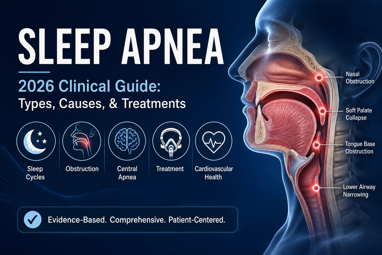
OSA is characterized by recurrent episodes of partial or complete upper airway collapse during sleep despite ongoing respiratory effort. The pathophysiology involves a combination of anatomical factors (narrowed airway, obesity, craniofacial structure) and neural factors (reduced pharyngeal dilator muscle activity during sleep, impaired airway reflexes).
Pathophysiology of OSA
Anatomical predisposition: The upper airway is a collapsible tube bounded by the soft palate, tongue, and lateral pharyngeal walls. Obesity increases fat deposition in the pharyngeal region, narrowing the airway. Craniofacial features including retrognathia (recessed mandible), micrognathia, and high-arched palate predispose to airway collapse.
Sleep-related muscle relaxation: During REM sleep, pharyngeal dilator muscles (genioglossus, tensor palatini) relax, reducing airway patency. In susceptible individuals, this relaxation allows airway collapse, initiating apnea. Arousal from sleep restores airway patency, but the cycle repeats throughout the night.
Ventilatory control instability: Impaired chemoreceptor sensitivity leads to oscillatory breathing patterns with periodic over- and under-ventilation, contributing to cycling apnea and hyperpnea.
III. Central Sleep Apnea (CSA): The Drive Failure
CSA results from absence of respiratory drive from the brainstem, with no respiratory effort during apneic episodes. It accounts for approximately 10-15% of sleep apnea cases.
Types of CSA:
Cheyne-Stokes respiration (CSR): Cyclic crescendo-decrescendo breathing pattern with central apneas during the nadir. Associated with heart failure, stroke, and renal failure. Characterized by prolonged circulation time and hyperventilation during the crescendo phase.
Primary CSA: Idiopathic central sleep apnea in the absence of cardiopulmonary or neurologic disease. Less common, often associated with altitude or opioid use.
Opioid-induced CSA: Chronic opioid use depresses central respiratory drive, causing irregular breathing patterns, central apneas, and ataxic breathing. Methadone and long-acting opioids are most commonly implicated.
Treatment-emergent CSA (Complex sleep apnea): New or worsening CSA occurring after CPAP initiation for OSA. Affects approximately 5-15% of OSA patients. Management often requires adaptive servo-ventilation (ASV).
CSA should be suspected in patients with: witnessed apneas without respiratory effort (no abdominal or thoracic movement), a history of heart failure (LVEF <45%), stroke, chronic opioid use, or Cheyne-Stokes breathing pattern on polysomnography. In-laboratory PSG is required for definitive diagnosis.
IV. Risk Factors for Sleep Apnea
| Risk Factor | Relative Risk | Mechanism |
|---|---|---|
| Obesity (BMI >30) | 4-5x | Fat deposition in pharyngeal tissues, reduced airway caliber |
| Male sex | 2-3x | Higher rates of obesity, different fat distribution |
| Aging (>60 years) | 3-4x | Increased upper airway collapsibility, reduced muscle tone |
| Neck circumference >17″ (men) or >16″ (women) | 4x | Direct marker of upper airway adiposity |
| Family history of OSA | 2x | Genetic predisposition, shared craniofacial features |
| Smoking | 1.5-2x | Airway inflammation, increased mucosal edema |
| Alcohol/sedative use | 2x | Increased pharyngeal muscle relaxation during sleep |
| Menopause (women) | 2x | Loss of hormonal protective effects on airway patency |
V. Clinical Presentation of Sleep Apnea
Nocturnal Symptoms
- Loud, habitual snoring: Often the first sign reported by bed partner. Snoring may be interrupted by pauses in breathing followed by gasping or choking sounds.
- Witnessed apneas: Bed partner observes breathing pauses lasting 10 seconds to over 1 minute, often followed by gasping or arousal.
- Choking or gasping: Awakening with sensation of breathlessness, often accompanied by palpitations or sweating.
- Nocturia: Frequent nighttime urination (≥2 times per night) due to increased sympathetic activity and reduced ADH secretion.
- Restless sleep: Frequent awakenings, tossing and turning, sometimes with night terrors or parasomnias.
Daytime Symptoms
- Excessive daytime sleepiness (EDS): Inappropriate sleepiness during waking hours, falling asleep while reading, watching television, driving, or in meetings. Epworth Sleepiness Scale score >10.
- Morning headaches: CO₂-mediated cerebral vasodilation, typically frontal or occipital, resolving within 1-2 hours after waking.
- Impaired cognition: Reduced attention, executive dysfunction, memory impairment, and slowed processing speed.
- Mood disturbances: Irritability, depression (30-40% of OSA patients), and anxiety.
- Reduced libido and erectile dysfunction: Hypoxemia and sleep fragmentation affecting hypothalamic-pituitary-gonadal axis.
• Witnessed apnea episodes lasting >30 seconds
• Severe daytime sleepiness with risk of falling asleep while driving
• Nocturnal angina or cardiac arrhythmias
• Severe morning headaches with hypertension
• Signs of heart failure or pulmonary hypertension
VI. Diagnostic Approach to Sleep Apnea
Polysomnography (PSG) — In-Laboratory Sleep Study
PSG is the gold standard for sleep apnea diagnosis. It monitors multiple physiological parameters including electroencephalogram (EEG), electrooculogram (EOG), electromyogram (EMG), electrocardiogram (ECG), airflow (nasal pressure, thermistor), respiratory effort (thoracic and abdominal inductance plethysmography), oxygen saturation (pulse oximetry), and body position. PSG is required for suspected CSA, heart failure, COPD, or when HSAT results are inconclusive.
Apnea-Hypopnea Index (AHI): The number of apneic and hypopneic events per hour of sleep. Normal: <5. Mild: 5-15. Moderate: 15-30. Severe: >30. AHI is the primary metric for diagnosis and severity classification.
Home Sleep Apnea Testing (HSAT)
HSAT is appropriate for patients with high pretest probability of moderate-severe OSA (STOP-BANG score ≥3, high BMI, witnessed apneas) without significant comorbidities. HSAT typically monitors respiratory effort, airflow, oxygen saturation, and heart rate. It is not recommended for patients with suspected CSA, insomnia, or significant cardiopulmonary disease.
STOP-BANG is an 8-item validated screening tool for OSA risk:
Snoring (loud enough to be heard through closed door)
Tiredness (daytime sleepiness or fatigue)
Observed apneas (breathing pauses during sleep)
Pressure (high blood pressure or treated hypertension)
BMI >35 kg/m²
Age >50 years
Neck circumference >40 cm (men) or >37 cm (women)
Gender (male)
Score 0-2: Low risk; 3-4: Moderate risk; 5-8: High risk.
VII. Evidence-Based Treatment of Sleep Apnea
Positive Airway Pressure (PAP) Therapy — First-Line Treatment
CPAP (Continuous Positive Airway Pressure): Delivers constant pressure throughout the respiratory cycle, maintaining upper airway patency. CPAP is the gold standard treatment for moderate-severe OSA and is effective in reducing AHI to <5, eliminating snoring, and improving sleep quality and daytime symptoms. Adherence is the key challenge — patients should use CPAP at least 4 hours per night for 70% of nights to derive benefit.
BiPAP (Bilevel Positive Airway Pressure): Provides higher inspiratory pressure (IPAP) and lower expiratory pressure (EPAP), assisting ventilation. BiPAP is indicated for patients requiring high pressures, those with COPD overlap syndrome, or those with treatment-emergent CSA. Settings typically IPAP 10-18 cm H₂O, EPAP 4-10 cm H₂O.
Adaptive Servo-Ventilation (ASV): Advanced device that adjusts pressure support to stabilize ventilation, indicated for CSA, Cheyne-Stokes respiration, and treatment-emergent CSA. Contraindicated in patients with severe heart failure with reduced EF (<45%) based on the SERVE-HF trial showing increased mortality.
Oral Appliances (Mandibular Advancement Devices)
Oral appliances are alternatives to PAP for patients who cannot tolerate CPAP or for mild-moderate OSA. The device holds the mandible forward during sleep, increasing upper airway caliber. Custom-fitted devices are more effective than over-the-counter or boil-and-bite devices. Efficacy is variable — AHI reduction is approximately 50% in responders.
Positional Therapy
Approximately 50-60% of OSA patients have positional OSA, where AHI is significantly higher in supine position. Positional therapy (sleeping on the side) is effective for patients with positional OSA and AHI <20. Methods include wearable devices, positional alarms, and specialized pillows.
Weight Loss and Lifestyle Modification
Weight loss of 10-15% of total body weight improves AHI by 30-50% and may cure OSA in some patients. Bariatric surgery can be curative for severely obese patients (BMI >40) with OSA. Additional lifestyle modifications include avoiding alcohol within 4 hours of bedtime, avoiding sedatives, and treating GERD.
Surgical Interventions
Surgery is considered for patients with severe OSA who fail or refuse PAP therapy and have surgically correctable anatomical abnormalities:
- UPPP (Uvulopalatopharyngoplasty): Removes excess soft palate and pharyngeal tissue. Success rate ~40-50%.
- Maxillomandibular advancement (MMA): Advances the upper and lower jaw, expanding the posterior airway space. Success rate ~80-90% but carries significant morbidity.
- Hypoglossal nerve stimulation (Inspire): Implantable device that stimulates the hypoglossal nerve during inspiration, advancing the tongue and opening the airway. Indicated for patients with BMI <32, AHI 15-65, and who have failed CPAP.
VIII. Interactive Clinical Tools
📊 STOP-BANG Sleep Apnea Risk Calculator
Validated 8-item screening tool for obstructive sleep apnea risk assessment.
Optimize Your Sleep Environment
Proper sleep positioning and ergonomic support can improve airway patency and reduce snoring. Ergonomic pillows and sleep systems maintain optimal head and neck alignment during sleep.
Explore Sleep Positioning Solutions →These statements have not been evaluated by the FDA. Individual results vary. Always consult your physician.
IX. Frequently Asked Questions
Sleep apnea is a sleep-related breathing disorder characterized by repeated episodes of complete (apnea) or partial (hypopnea) upper airway obstruction during sleep, leading to intermittent hypoxemia, arousal, and daytime sleepiness. It affects approximately 25% of middle-aged men and 10% of middle-aged women.
OSA results from upper airway collapse despite respiratory effort. CSA results from absence of respiratory drive from the brainstem, with no respiratory effort during apneic episodes. CSA is less common and associated with heart failure, stroke, or opioid use.
Normal AHI is <5 events/hour. Mild OSA: 5-15. Moderate: 15-30. Severe: >30. AHI guides treatment decisions. AHI >15 with symptoms, or >30 regardless, generally warrants treatment.
STOP-BANG is an 8-item screening tool: Snoring, Tiredness, Observed apneas, Pressure (hypertension), BMI >35, Age >50, Neck circumference >40 cm (men) or >37 cm (women), Gender (male). Score 0-2: low risk; 3-4: moderate; 5-8: high risk for OSA.
First-line treatment for moderate-severe OSA is positive airway pressure (CPAP or BiPAP). Alternatives include oral appliances, positional therapy, weight loss, and upper airway surgery. Lifestyle modifications include weight loss, positional sleep, and avoiding alcohol/sedatives before bed.
Untreated sleep apnea is associated with hypertension, cardiovascular disease (myocardial infarction, stroke, atrial fibrillation), heart failure, type 2 diabetes, cognitive impairment, depression, motor vehicle accidents, and increased all-cause mortality. CPAP therapy reduces cardiovascular mortality by 40-60%.
OSA is typically a chronic condition that is managed rather than cured. Weight loss can cure OSA in a minority of patients (10-20% with significant weight loss). Surgery may be curative in select patients but carries significant morbidity. CPAP effectively eliminates apneic events during use.
Diagnosis requires polysomnography (in-laboratory sleep study) or home sleep apnea testing (HSAT). HSAT is appropriate for patients with high pretest probability of moderate-severe OSA without significant comorbidities. PSG is recommended for suspected CSA, heart failure, or when HSAT results are inconclusive.
Support Respiratory and Sleep Health
Targeted nutritional support — including magnesium, glycine, and antioxidant formulations — may help support sleep quality and respiratory function. Consult your physician before starting any supplement regimen.
Explore Sleep Support Formulas →These statements have not been evaluated by the FDA. This product is not intended to diagnose, treat, cure, or prevent any disease. Individual results vary.
Leave a Reply